题目
AVBS2001 (ND) Prep for Microscopy Prac 2 in week 3 (1%): Disorders of Growth and Circulatory
多重下拉选择题
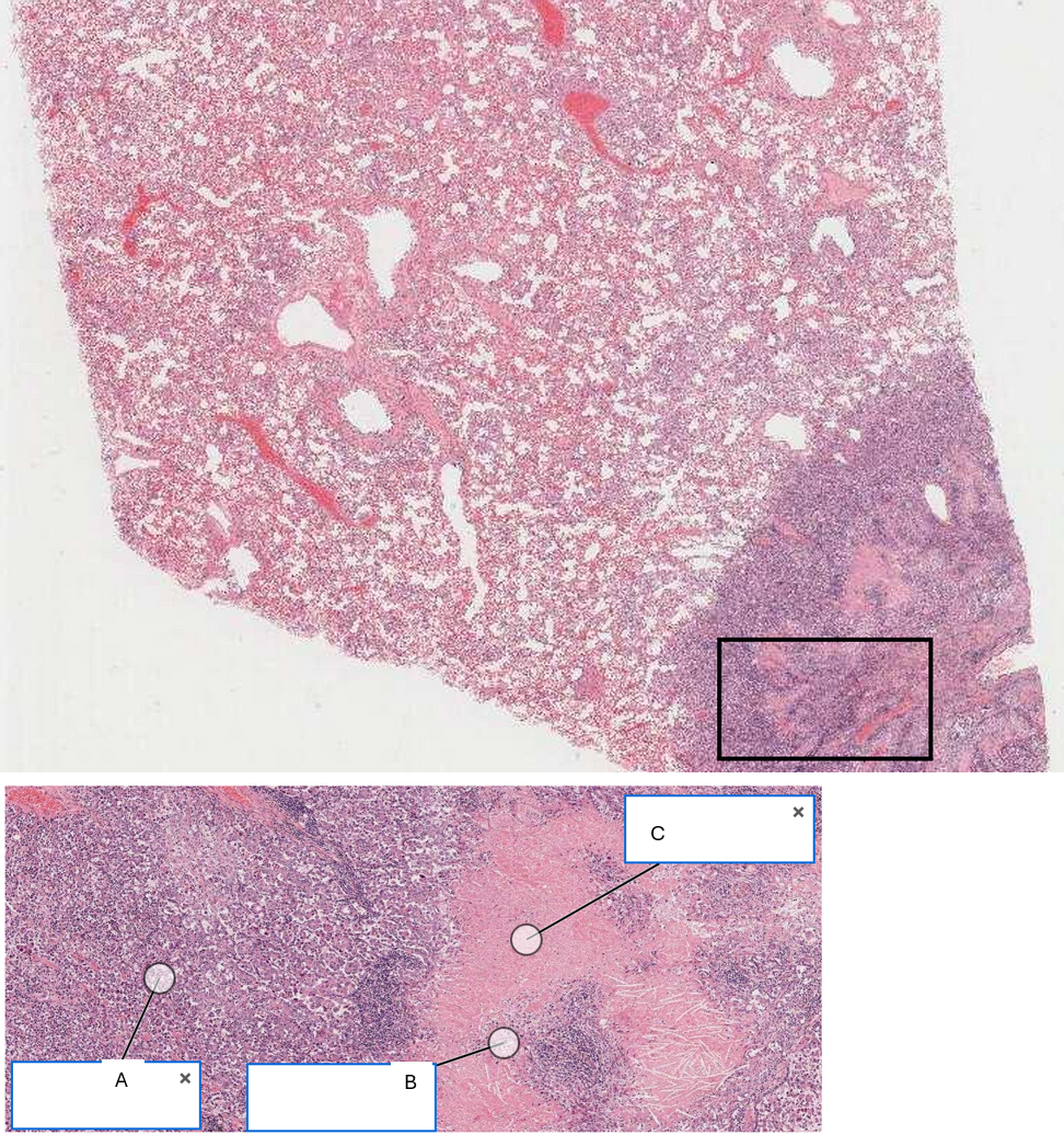
The remainder of the section appears more dense. Here some areas are highly cellular, while others appear as amorphous pink material with some nuclear debris. There is also marked distension of blood vessels. Name the labels below: A: Highly cellular area , B: Nuclear fragments/ debris , C: Amorphous pink material
查看解析
标准答案
Please login to view
思路分析
To approach this labeling task, I’ll evaluate what each description typically corresponds to in a histology image showing a mixture of cellular and acellular areas.
Option A: 'Highly cellular area' – This label should correspond to regions with a high density of cells and numerous nuclei packed closely together. In many slides, such areas appear darkened due to ma......Login to view full explanation登录即可查看完整答案
我们收录了全球超50000道考试原题与详细解析,现在登录,立即获得答案。
类似问题
The best place to view the primary pathological process in action is generally on the edge of the lesion- in the middle it will often be confused by secondary changes, such as necrosis and haemorrhage. Normal lung to left of image. On right note infiltration of alveolar spaces by cells. Note different sizes and shapes of cells (pleiomorphism), some binucleated cells, some joined and others not (degrees of de-differentiation) Name the labels for the section below: A: Cells within alveolar spaces , B: Cells forming a line , C: Alveolar wall .
In one area, there is a raised, dome-like lesion. The mass is well demarcated and doesn’t extend into the subcutis. The lesion is covered by a thin layer of epidermis, which has become ulcerated and shows superficial infiltration by individual round cells with a lobulated nucleus (neutrophils) and plump spindle-shaped mesenchymal cells (likely fibroblasts and vascular endothelial cells in repair). Label the features: A: Collagen Fibres , B: Neutrophils , C: Mesenchymal cells (fibroblasts or endothelial cells)
Time for an abnormal section 001(1) Click the link Links to an external site. to activate your virtual microscopy page. You should see your two normal liver sections, which you can use for comparison. Below them, click on section 001(1) and examine the section. Below are some fragments of text that, together, make up a description for this section. Indicate if you think each are architecture, constituent components, non-constituent components, or interpretation: 1. The section shows a piece of liver. Architecture 2. Central veins, and portal triads can be seen clearly in normal lobular arrangement Architecture 3. The portal triads consist of a bile duct, portal vein and hepatic arteriole though at times these features are indistinct Constituent components 4. The central veins are clearly visible Constituent components 5. The hepatic cords and sinusoids are not clear Constituent components 6. There is widespread vacuolation of the cytoplasm of the hepatocytes. This ranges from many small vacuoles within an eosinophilic cytoplasm in hepatocytes near the central vein; to large vacuoles completely filling and distending the cytoplasm and pushing the small dense nucleus to the side in hepatocytes closer to the portal triads. Constituent components 7. Within the connective tissue of the portal triads there are many individual cells with a small dense round basophilic nucleus and little cytoplasm (lymphocytes). Non-constituent components 8. Widespread periportal hepatic lipidosis Interpretation 9. Reversible cell injury and mild periportal inflammation Interpretation
The image below shows some brown material within H&E stained cells. Can you identify the material?
更多留学生实用工具
希望你的学习变得更简单
加入我们,立即解锁 海量真题 与 独家解析,让复习快人一步!