题目
AVBS2001 (ND) Prep for Microscopy Prac 2 in week 3 (1%): Disorders of Growth and Circulatory
多重下拉选择题
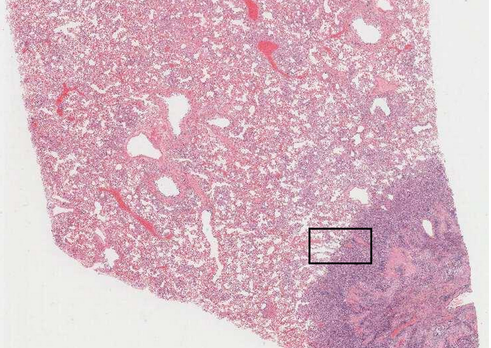
The best place to view the primary pathological process in action is generally on the edge of the lesion- in the middle it will often be confused by secondary changes, such as necrosis and haemorrhage. Normal lung to left of image. On right note infiltration of alveolar spaces by cells. Note different sizes and shapes of cells (pleiomorphism), some binucleated cells, some joined and others not (degrees of de-differentiation) Name the labels for the section below: A: Alveolar wall , B: Cells forming a line , C: Cells within alveolar spaces .
查看解析
标准答案
Please login to view
思路分析
To approach this question, we first restate what is being asked and what each label is supposed to identify in the histology image.
- A: Alveolar wall
- B: Cells forming a line
- C: Cells within alveolar spaces
Option 1 (A: Alveolar wall) analysis:
The alveolar walls (septae) are the structural partitions between adjacent alveoli. In many lung pathology images, primary processes such as inflammatory or neoplastic changes can be most evident at the edge of a lesion where the architecture is disrupted but not yet overwhelmed by secondary changes. If the box in the image is highlighting tissue at the thin connec......Login to view full explanation登录即可查看完整答案
我们收录了全球超50000道考试原题与详细解析,现在登录,立即获得答案。
类似问题
Question at position 20 Which one of the following requires magnification to study? Histology Regional anatomy Gross anatomy Surface anatomy
In the colon, the muscularis externa is primarily composed of Blank 1 Question 17[select: , skeletal muscle, smooth muscle, dense connective tissue, lamina propria] , while the mucosal epithelium consists of Blank 2 Question 17[select: , stratified cuboidal, simple squamous, simple columnar, stratified squamous] cells.
In one area, there is a raised, dome-like lesion. The mass is well demarcated and doesn’t extend into the subcutis. The lesion is covered by a thin layer of epidermis, which has become ulcerated and shows superficial infiltration by individual round cells with a lobulated nucleus (neutrophils) and plump spindle-shaped mesenchymal cells (likely fibroblasts and vascular endothelial cells in repair). Label the features: A: Mesenchymal cells (fibroblasts or endothelial cells) , B: Neutrophils , C: Collagen Fibres
Name the endocrine organ featured in this micrograph (histological image)
更多留学生实用工具
希望你的学习变得更简单
加入我们,立即解锁 海量真题 与 独家解析,让复习快人一步!