Questions
AVBS2001 (ND) Prep for Microscopy Prac 2 in week 3 (1%): Disorders of Growth and Circulatory
Multiple dropdown selections
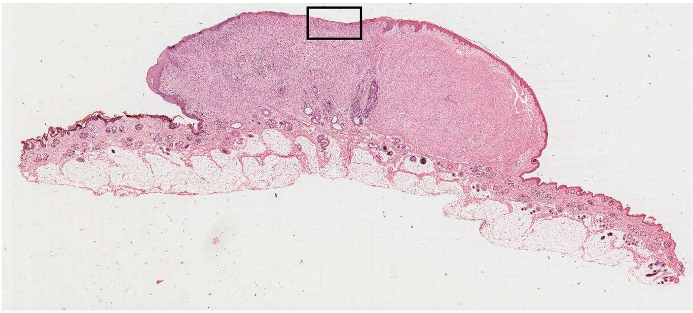
In one area, there is a raised, dome-like lesion. The mass is well demarcated and doesn’t extend into the subcutis. The lesion is covered by a thin layer of epidermis, which has become ulcerated and shows superficial infiltration by individual round cells with a lobulated nucleus (neutrophils) and plump spindle-shaped mesenchymal cells (likely fibroblasts and vascular endothelial cells in repair). Label the features: A: Mesenchymal cells (fibroblasts or endothelial cells) , B: Neutrophils , C: Collagen Fibres
View Explanation
Verified Answer
Please login to view
Step-by-Step Analysis
The task asks us to label the features A, B, and C on the histology image based on what is visible in the lesion.
Option A: Mesenchymal cells (fibroblasts or endothelial cells)
- In a healing or repair setting, mesenchymal cells such as fibroblasts and vascular endothelial cells are prominent components of granulation tissue. They appear as spindle-shaped cells within the connective tissue and are responsible for laying down new extracellular matrix and forming new blood vessels. In the image, the presence of plum......Login to view full explanationLog in for full answers
We've collected over 50,000 authentic exam questions and detailed explanations from around the globe. Log in now and get instant access to the answers!
Similar Questions
Question at position 20 Which one of the following requires magnification to study? Histology Regional anatomy Gross anatomy Surface anatomy
In the colon, the muscularis externa is primarily composed of Blank 1 Question 17[select: , skeletal muscle, smooth muscle, dense connective tissue, lamina propria] , while the mucosal epithelium consists of Blank 2 Question 17[select: , stratified cuboidal, simple squamous, simple columnar, stratified squamous] cells.
The best place to view the primary pathological process in action is generally on the edge of the lesion- in the middle it will often be confused by secondary changes, such as necrosis and haemorrhage. Normal lung to left of image. On right note infiltration of alveolar spaces by cells. Note different sizes and shapes of cells (pleiomorphism), some binucleated cells, some joined and others not (degrees of de-differentiation) Name the labels for the section below: A: Alveolar wall , B: Cells forming a line , C: Cells within alveolar spaces .
Name the endocrine organ featured in this micrograph (histological image)
More Practical Tools for Students Powered by AI Study Helper
Making Your Study Simpler
Join us and instantly unlock extensive past papers & exclusive solutions to get a head start on your studies!