Questions
BIOL 271
Single choice
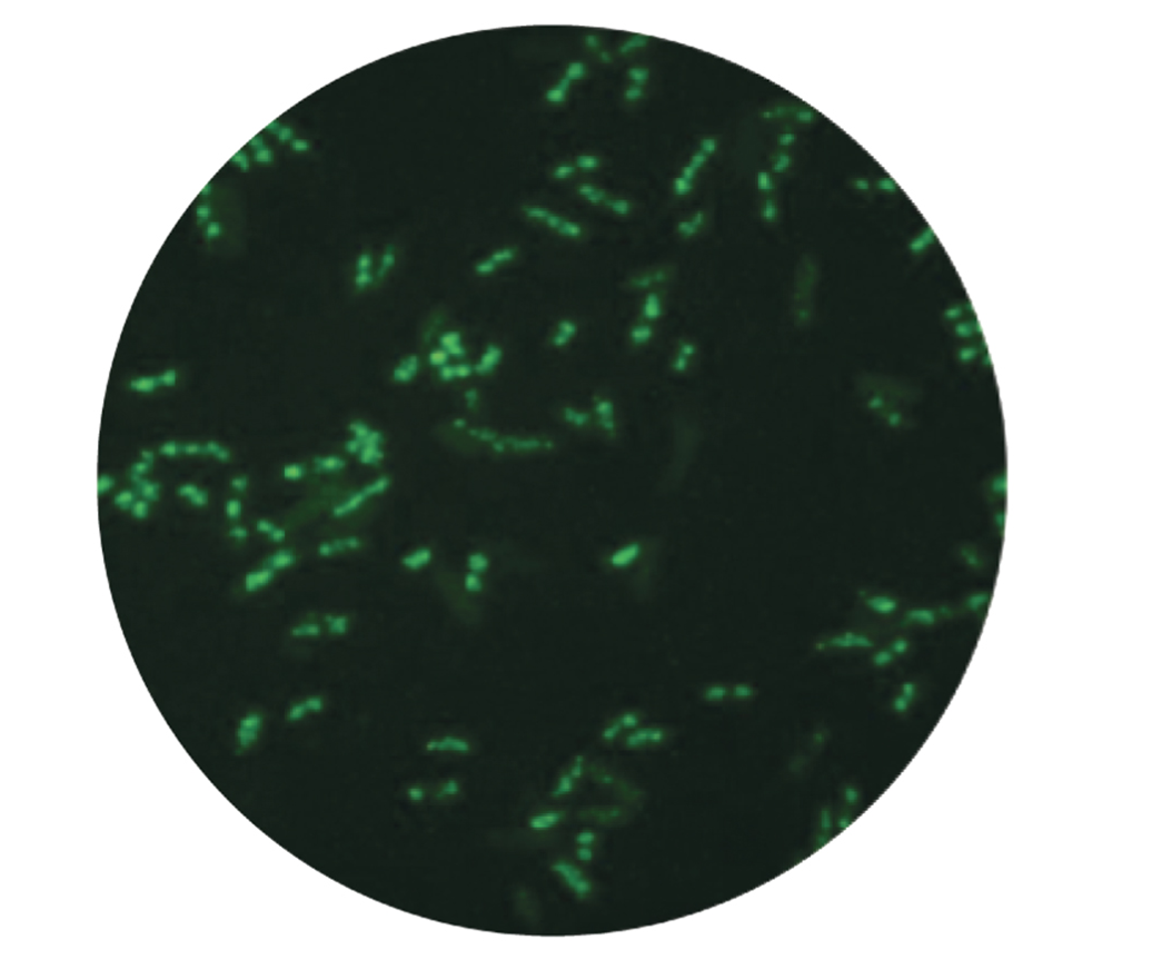
Select the type of microscopy used to generate the image:
View Explanation
Verified Answer
Please login to view
Step-by-Step Analysis
The prompt asks us to identify the type of microscopy used to generate the image. The image shows bright green punctate structures against a dark background, which is a hallmark of fluorescence-based detection where fluorophores emit light at a specific wavelength when excited.
First, consider fluorescence microscopy: it uses fluorescent labels to tag specific components, pro......Login to view full explanationLog in for full answers
We've collected over 50,000 authentic exam questions and detailed explanations from around the globe. Log in now and get instant access to the answers!
Similar Questions
In confocal microscopy, fluorescence occurs due to
This type of microscope is best suited for visualizing GFP, RFP, and YFP proteins.
True or False? Ultraviolet wavelengths of light are required to visualise plasma membranes of cells. (1 mark)
In a consumer society, many adults channel creativity into buying things
More Practical Tools for Students Powered by AI Study Helper
Making Your Study Simpler
Join us and instantly unlock extensive past papers & exclusive solutions to get a head start on your studies!